Down Sendromu Nedir?

Down Sendromu Nedir?

Genetik bir durumdur ve 21. kromozom çiftinin fazlalığı nedeniyle ortaya çıkar. Bu fazlalık, zihinsel ve fiziksel gelişim üzerinde etkili olur. down syndrome yaşayan bireylerde tipik yüz hatları, kas gevşekliği ve öğrenme güçlükleri görülebilir. Kalp ve sindirim sistemi sorunları da yaygındır. Erken müdahale ve özel eğitimle bu bireylerin yaşam kalitesi artırılabilir.

Down Sendromu Belirtileri Nelerdir?

Down Sendromu Nedir?Burun kökü basık ve yüzde genel bir yassı görünüm vardır. Gözler yukarı doğru çekik, badem şekillidir. Gözlerde epikantal kıvrım (göz iç köşelerinde deri kıvrımı) olabilir. Burun kökü basık, burun kısa ve geniş olabilir. down syndrome olan bireylerde ağız küçük olabilir ve dil genellikle dışarıda durur. Boyun kısa ve geniş bir görünüme sahip olabilir. Avuç içindeki çizgiler normalden farklı olarak tek bir çizgi şeklinde olabilir.

Kulaklar normalden daha küçük ve düşük yerleşimli olabilir. Bebeklik döneminde kaslar zayıf olabilir, bu da hareket ve motor gelişimi etkileyebilir. Eller ve parmaklar kısa ve kalın olabilir. Ayrıca serçe parmağı içe doğru kıvrık olabilir. Zihinsel gelişimde bazı gecikmeler olabilir. Bu da öğrenme ve dil gelişimini etkileyebilir. Doğuştan kalp anomalileri sık görülebilir. Genellikle yaşlarına göre daha kısa boylu olabilirler. Bu belirtiler kişiden kişiye farklılık gösterebilir. Her down syndrome yaşayan birey bu belirtilerin hepsini taşımayabilir.

Down Sendromu Nasıl Teşhis Edilir?

İkili tarama testi gebeliğin 11-14. haftalarında yapılır. Bu test, annenin kanındaki bazı hormonların seviyelerini ve ultrason ile ölçülen ense kalınlığını analiz eder. down syndrome riski hakkında bilgi verir. Üçlü veya dörtlü tarama testi, gebeliğin 15-20. haftalarında yapılır. Annenin kanındaki farklı hormon seviyelerini inceler ve risk hakkında bilgi verir. Gebeliğin 15-20. haftalarında amniyotik sıvıdan örnek alınarak bebeğin kromozomları incelenir. Kesin bir tanı sağlar. Koryon villus biyopsisi (CVS), gebeliğin 10-13. haftalarında yapılır.

Bu testte plasenta dokusundan örnek alınır ve kromozom analizi yapılır. Kordosentez, gebeliğin ilerleyen haftalarında (20. haftadan sonra) bebekten kan alınarak yapılan bir testtir. Kromozom anomalileri bu testle de tespit edilebilir. Kromozom analizinde (Karyotip), bebeğin kan örneği alınarak kromozom yapısı incelenir. Down sendromu tanısı bu yöntemle kesinleştirilir. down syndrome teşhisi konulursa, aileye genetik danışmanlık verilir. Durum hakkında bilgilendirilir. bebek için uygun tedavi ve destek planları oluşturulur.

Down Sendromu Neden Olur?

Down Sendromu Nedir?En yaygın nedeni, hücre bölünmesi sırasında 21. kromozomun fazladan bir kopyasının oluşmasıdır. Bu, 21. kromozomun üç kopyasına sahip olma durumudur (Trizomi 21). İleri anne yaşı, down syndrome riskini artıran faktörlerden biridir. Özellikle 35 yaş ve üzeri annelerde risk artmaktadır. Nadiren, kalıtsal olabilir. Translokasyon adı verilen bir genetik değişiklik sonucu, ebeveynden çocuklarına aktarılır.

Döllenmeden sonraki hücre bölünmesi sırasında meydana gelen hatalar da yol açabilir. Bu durum, hücrelerin bölünme sürecinde genetik materyalin hatalı bir şekilde dağılmasıyla ortaya çıkar. Nadiren, kişinin bazı hücrelerinde normal kromozom sayısı (46) varken, diğer hücrelerde 47 kromozom bulunur. Bu durum, mosaic down syndrome olarak bilinir. Farklı hücrelerde farklı genetik yapılar oluşmasına neden olur.

Down Sendromu Tedavi Yöntemleri Nelerdir?

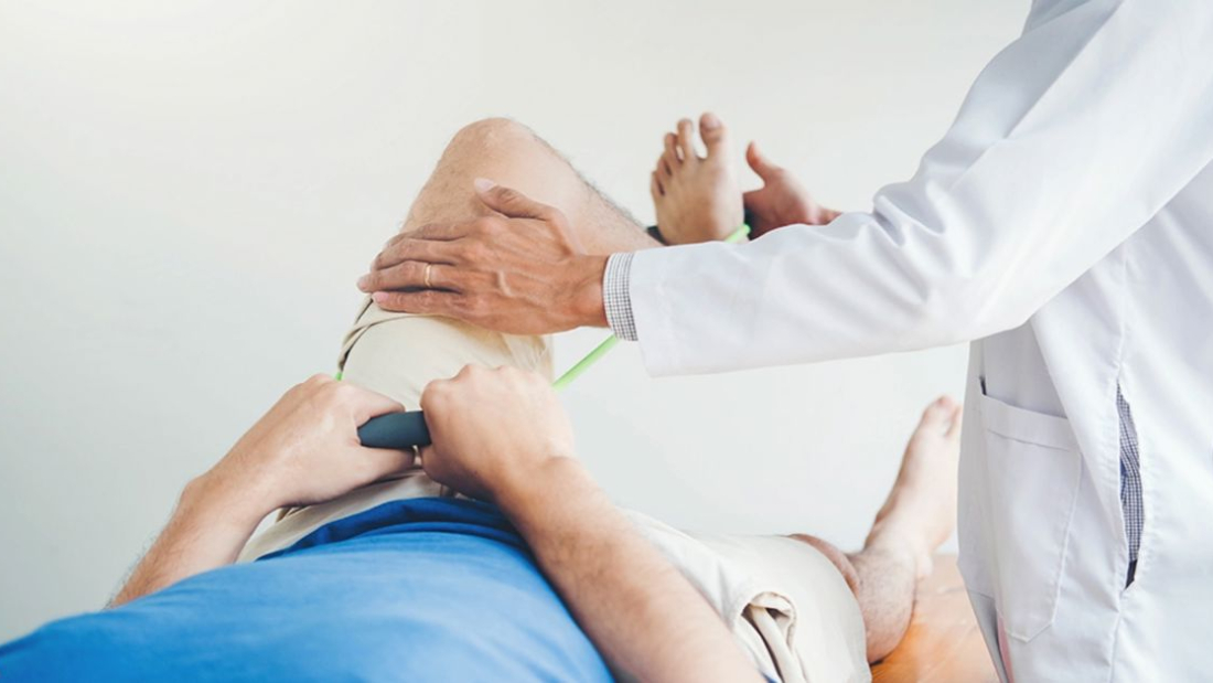
Bebeğin gelişimini desteklemek için fizyoterapi, dil terapisi ve işitme terapisi uygulanır. Özel eğitim programları ile bireyin öğrenme kapasiteleri artırılır. Kas tonusunu artırmak ve motor becerilerini geliştirmek için uygulanır. Dil gelişimini desteklemek ve iletişim becerilerini artırmak için kullanılır. Tiroid sorunları gibi eşlik eden sağlık problemleri için ilaçlar kullanılır. Aile ve birey için psikolojik danışmanlık hizmetleri sunulur. Sağlıklı bir yaşam için uygun beslenme programları uygulanır.

Pençe Parmak Hastalığı Nedir?

Pençe Parmak Hastalığı Nedir?

Parmakların sürekli bükülü bir şekilde kalmasına yol açan bir deformitedir. Pençe parmak hastalığı genellikle sinir ve kaslardaki zayıflık ya da hasar nedeniyle ortaya çıkar. El ve ayak parmaklarında görülebilir. Tedavi edilmezse hareket kısıtlılığına ve ağrıya neden olabilir. Fizik tedavi, ateller ve cerrahi yöntemler ile tedavi edilebilir. Erken tanı, ilerlemesini durdurmada önemlidir.

Pençe Parmak Hastalığı Belirtileri Nelerdir?

Pençe Parmak Hastalığı Nedir?Ayak parmakları, özellikle ikinci, üçüncü ve dördüncü parmaklar bükülerek bir pençe görünümü alır. Parmaklar yukarı doğru kalkar ve eklemler bükülü kalır. Parmaklardaki bükülme, özellikle yürürken veya ayakkabı giyerken ağrıya neden olabilir. Bu ağrı, genellikle eklemlerde ve parmak uçlarında hissedilir. Parmakların bükülü durması, ayak parmaklarının uçlarında ve eklemlerde sürtünmeye neden olur, bu da nasırların oluşmasına yol açabilir. Ayrıca, ayak mantarına eğilim artabilir. Pençe parmak hastalığı, ayağın dengesi üzerinde olumsuz etkiler yaparak yürümeyi zorlaştırabilir. Zamanla parmaklardaki bükülme sertleşebilir ve bu durum, parmakların normale döndürülmesini zorlaştırabilir. Genellikle ayakkabı seçimi, kas zayıflığı veya nörolojik problemler nedeniyle gelişebilir. Tedavi egzersiz, ortopedik ayakkabılar veya ileri vakalarda cerrahi müdahale ile yapılır.

Pençe Parmak Hastalığı Neden Olur?

Sinirlerde meydana gelen hasarlar, pençe parmak hastalığı yaşanmasına yol açabilir. Özellikle periferik sinir yaralanmaları ve nörolojik hastalıklar bu duruma neden olur. El ve ayak parmaklarındaki kasların zayıflaması veya dengesiz çalışması, sebep olabilir. Bu durum, kas distrofisi gibi kas hastalıklarında da görülebilir. Diyabet hastalarında sıkça görülen nöropati (sinir hasarı), yol açabilir. Romatoid artrit gibi inflamatuar romatizmal hastalıklar, eklemlerde yapısal bozukluklara neden olur. Bu durumun gelişimine neden olur. Parmaklarda meydana gelen travma veya kırıklar, doğru şekilde tedavi edilmediğinde neden olur. Bazı bireyler doğuştan sahip olabilir. Pençe parmak hastalığı tedavisi, altta yatan nedenin belirlenmesi ile gerçekleştirilir.

Pençe Parmak Hastalığı Tedavisi Nasıl Yapılır?

İlk olarak, bir ortopedi uzmanı tarafından detaylı bir fiziksel muayene gerçekleştirilir. Röntgen gibi görüntüleme yöntemleri kullanılarak, deformitenin şiddeti ve nedenleri belirlenir. Hafif vakalarda, ayak parmağı için özel ayakkabı, parmak destekleri ve egzersiz önerilir. Bu yöntemler, kasları güçlendirip parmakları düzeltmeyi hedefler. Aynı zamanda, ağrı ve şişliği azaltmak için anti-inflamatuar ilaçlar reçete edilebilir. İleri seviye vakalarda ise cerrahi müdahale gerekebilir. Bu cerrahide, tendonların gevşetilmesi, eklemlerin düzeltilmesi işlemleri yapılabilir. Pençe parmak hastalığı tedavi yöntemi, hastalığın ilerlemesine ve kişinin yaşam kalitesine bağlı olarak belirlenir. Erken müdahale ile konservatif tedavi genellikle başarılı olabilir. Ancak daha ileri durumlar için cerrahi seçenekler değerlendirilmelidir.

Tedavi Sonrası Dikkat Edilmesi Gerekenler Nelerdir?

Pençe Parmak Hastalığı Nedir?Ameliyat veya konservatif tedavi sonrası, kas gücünü ve esnekliği geri kazanmak için fizik tedavi önerilir. Egzersizler düzenli olarak yapılmalı ve doktorun talimatlarına uyulmalıdır. Operasyon sonrası dikişlerin bakımı hijyenik bir şekilde yapılmalı. Doktorun önerdiği pansumanlar düzenli olarak uygulanmalıdır. Enfeksiyon belirtilerine karşı dikkatli olunmalıdır. Parmakları zorlamadan dinlenmek, iyileşme sürecinde önemlidir. Ağır aktivitelerden kaçınmalı ve iyileşme tamamlanana kadar parmaklar korunmalıdır. Düzenli doktor kontrolleri, iyileşme sürecinin takibi açısından önemlidir. Herhangi bir komplikasyon durumunda erken müdahale sağlanabilir. İyileşme sürecinde sağlıklı bir beslenme ve yeterli su tüketimi, vücudun onarım sürecine katkıda bulunacaktır. Bu noktalara dikkat edilmesi, tedavi sonuçlarını olumlu yönde etkileyerek daha hızlı bir iyileşme sağlayabilir.

Stres Kırığı Nedir?

Stres Kırığı Nedir?

Tekrarlayan küçük travmalar veya aşırı kullanım sonucu oluşan ince kemik çatlağıdır. Stres kırığı genellikle ayak, bacak veya kalçada ortaya çıkar ve koşucular, askerler veya dansçılarda sık görülür. Zamanla gelişen ağrı, başlangıçta hafif olup, dinlenmeyle azalır ancak devam ettikçe daha şiddetli hale gelebilir. Tedavi edilmezse, daha büyük kırıklara yol açabilir.

Stres Kırığı Belirtileri Nelerdir?

Stres Kırığı Nedir?
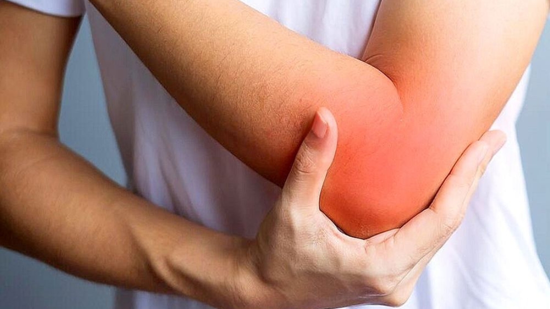
Etkilenen bölgede ilk başta hafif olan ve zamanla şiddetlenen bir ağrı hissedilir. Ağrı, genellikle aktiviteler sırasında artar ve dinlenmeyle azalır. Kırığın bulunduğu bölgede hafif bir şişlik meydana gelir. Kemik üzerindeki baskıya karşı hassasiyet artar. Bu bölgede dokunulduğunda ağrı hissedilir. Nadiren de olsa ciltte hafif renk değişiklikleri gözlemlenebilir. Zamanla yaralanma bölgesindeki ağrı, günlük aktiviteleri gerçekleştirmeyi zorlaştırabilir. Hareket kısıtlılığı yaratabilir. Stres kırığı belirtisi fark edildiğinde, profesyonel bir değerlendirme ve tedavi gerekir.

Stres Kırığı Neden Olur?

Ani artan ya da aşırı tekrarlanan egzersizler, kemiklerde mikro hasarlara yol açarak neden olur. Özellikle uzun mesafe koşucuları, dansçılar ve askeri personel bu durumdan sıklıkla etkilenir. Kemiklerin kendini yenilemesi ve iyileşmesi için dinlenme önemlidir. Yetersiz dinlenme, kemiklerin iyileşme sürecini engelleyerek stres kırığı yaşanmasına zemin hazırlar. Osteoporoz veya yetersiz kalsiyum ve D vitamini alımı gibi faktörler kemik yoğunluğunu azaltır. Kemikleri daha kırılgan hale getirebilir.

Spor ayakkabılarının yanlış seçilmesi veya aşınmış ayakkabıların kullanılması, ayak ve bacak kemiklerine aşırı yük bindirerek yol açar. Düz tabanlık, bacak uzunluğu farkı veya yanlış koşu tekniği gibi sorunlar, vücudun belirli bölgelerine aşırı yük binmesine neden olur. Bu durumun yaşanmasına neden olabilir. Normalde yapılan aktivitelerde ani bir artış yaşanması, kemiklere aşırı yük bindirerek oluşumunu tetikler. Yetersiz beslenme, özellikle düşük enerji alımı, kemik sağlığını olumsuz etkileyebilir ve stres kırığı riskini artırabilir.

Stres Kırığı Nasıl Teşhis Edilir?

Doktor, ağrının ne zaman başladığını, hangi aktiviteler sırasında arttığını ve ağrının niteliğini sorgular. Ardından ağrılı bölgeyi muayene ederek, hassasiyet olup olmadığını kontrol eder. Erken dönem stres kırığı röntgende her zaman görünmeyebilir. Genellikle 2-3 hafta sonra kemikte değişiklikler gözlemlenebilir. Manyetik rezonans görüntüleme erken teşhisinde en hassas yöntemlerden biridir.

Yumuşak dokular ve kemik içindeki küçük çatlaklar detaylı bir şekilde görüntülenebilir. Kemik tarama, kemik dokusundaki aktif iyileşme süreçlerini tespit eder. Kemik taramaları, bu durumun olduğu bölgede artan kan akışını gösterir. Bilgisayarlı tomografi, kemik yapısının daha ayrıntılı görüntülenmesi gerektiğinde kullanılır. Özellikle karmaşık bölgelerdeki stres kırığı için tercih edilebilir. Erken teşhis ve tedavi, kırığın ilerlemesini önleyerek iyileşme sürecini hızlandırır.

Stres Kırığı Tedavi Yöntemleri Nelerdir?

Stres Kırığı Nedir?Stres kırığı olan bölgeyi iyileştirmek için birkaç hafta boyunca dinlenmek en temel tedavi yöntemidir. Etkilenen bölgeye aşırı yüklenmekten kaçınılmalıdır. Ağrıyı ve şişliği azaltmak için günde birkaç kez buz uygulaması yapılabilir. Ayak veya bacakta varsa, uygun destek sağlayan ayakkabılar veya özel ortopedik cihazlar kullanılır. Kasları güçlendirmek ve esnekliği artırmak amacıyla fizyoterapist eşliğinde egzersiz programı uygulanır.

İyileşme sürecinde, normal aktivitelere yavaş yavaş geri dönmek önemlidir. Tam iyileşmeden önce aşırı egzersizlerden kaçınılmalıdır. Kemik sağlığını desteklemek amacıyla kalsiyum ve D vitamini takviyesi önerilebilir. Daha ciddi vakalarda, alçı veya destek bandajı kullanılabilir. Nadiren, tedaviye yanıt vermeyen stres kırığı için cerrahi müdahale gerekebilir. Bu yöntemler kırığın ciddiyetine ve kişinin sağlık durumuna göre değişiklik gösterebilir.

Tennis Elbow Disease

Tennis Elbow Disease

It is a disorder that occurs as a result of excessive use of the muscles and tendons located on the outer part of the elbow. Tennis elbow disease, occurs due to repetitive movements of the wrist and hand. That's why it got its name from tennis players. However, it is common not only in tennis players but also in people who use their wrists frequently.

Symptoms of Tennis Elbow Disease

Tennis Elbow Disease

It is a disorder that occurs as a result of excessive use of the tendons located on the outside of the elbow. This disease occurs due to repetitive hand and wrist movements. It usually manifests itself with pain and tenderness on the outside of the elbow. Tennis elbow symptoms usually develop gradually and become more severe over time.

The most common symptom is pain on the outside of the elbow. This pain may be mild at first, but if left untreated, it can become more severe over time. The pain may increase with simple movements, such as shaking hands, turning a doorknob, or lifting an object. The pain becomes more noticeable during activities that use the wrist and forearm frequently.

Another important symptom is tenderness and discomfort on the outside of the elbow. Patients may feel pain when they touch or strain this area of the elbow. In some cases, a feeling of weakness in the elbow may also occur, which can make daily activities difficult for the person.

In addition to pain and tenderness, swelling and a slight increase in temperature in the elbow may also be observed. Rarely, limited movement in the elbow area may develop. These symptoms are more common in advanced cases of the disease. In addition, if left untreated, it can negatively affect daily life.

Tennis elbow disease It is important to see a specialist early when symptoms are noticed. Early diagnosis and appropriate treatment can stop the progression of the disease and speed up the recovery process.

Causes of Tennis Elbow Disease

Tennis elbow is a disease that develops due to excessive use of the muscles and tendons located on the outside of the elbow. This disorder occurs in repetitive hand and wrist movements. Although tennis elbow takes its name from the sport of tennis, the causes of this disease are not limited to sports.

The most common cause is repetitive movements. These movements cause the muscles and tendons to be constantly strained and eventually inflamed. This condition is especially triggered by repeated movements such as bending, lifting, squeezing and turning the wrist. It is also common in people who use hand tools.

In addition, using the mouse constantly, pressing the keyboard keys hard or typing for long periods of time are among the causes. In addition, sports activities with poor technique, working in the wrong position or improper equipment also cause it to occur.

Age is another factor. The risk of tennis elbow increases, especially in individuals between the ages of 30 and 50, as the flexibility of muscles and tendons decreases. This makes the muscles more susceptible to overuse and strain.

In conclusion, tennis elbow disease, mostly occurs as a result of repetitive movements and overuse. Therefore, it is important to regulate activities and consult a specialist when symptoms are felt.

Tennis Elbow Disease Treatment Methods

Tennis Elbow Disease

Tennis elbow is a disorder that occurs as a result of excessive use of the tendons located on the outside of the elbow. If this disease is not treated promptly and appropriately, it can negatively affect the daily lives of patients. Treatment methods usually vary depending on the severity of the disease and the general health of the patient.

The initial treatment approach begins with rest and activity modification. Patients should avoid repetitive movements to reduce pressure on the elbow. They should also take care to rest. During this process, applying cold can be effective in reducing pain and inflammation.

Physical therapy plays an important role in the treatment of tennis elbow. A physical therapist will provide the patient with specific stretching and strengthening exercises that will help strengthen the elbow muscles and tendons. These exercises should be performed carefully to reduce the risk of re-injury.

Medication is also a common treatment. Nonsteroidal anti-inflammatory drugs (NSAIDs) are effective in reducing pain and inflammation. In more severe cases, doctors may recommend corticosteroid injections. However, this treatment method is not recommended for long-term use.

Surgery may be considered when other treatments have failed. Surgery involves repairing or removing damaged tissue. Rehabilitation after surgery is important for a full recovery.

Tennis elbow disease treatment, early intervention and treatment methods prevent the progression of the disease. In addition, regular exercise and rest are also important in this process. This treatment approach can improve the quality of life of patients.

What is a Shoulder Fracture?

What is a Shoulder Fracture?

Omuz kırığı, omuz eklemini oluşturan kemiklerden birinin bütünlüğünün bozulmasıdır. En sık humerus kemiğinin üst kısmında görülür. Köprücük kemiği ve kürek kemiği de bu tabloya dâhil olabilir. Bu durumda omuz kırığı nedir? sorusu sıkça sorulur. Omuz eklemi oldukça hareketlidir. Bu nedenle travmalara karşı daha hassastır.

Omuz Kırığı Belirtileri Ve Nedenleri

What is a Shoulder Fracture?

Omuz bölgesi, kolun vücutla bağlantısını sağlayan karmaşık bir yapıya sahiptir. Günlük yaşamda sık kullanılan bu eklem, düşme ve çarpmalara karşı hassastır. Ani travmalar omuz çevresinde ciddi hasarlara yol açabilir. Özellikle kol üzerine düşmelerde ağrı hızla artar. Hareket kısıtlılığı kısa sürede fark edilir. Şişlik ve morarma da bu tabloya eşlik edebilir. Kişi kolunu doğal pozisyonunda tutmakta zorlanabilir.

Travma sonrası ortaya çıkan ağrı genellikle şiddetlidir ve hareketle artar. Shoulder fracture, çoğu zaman kolu kaldırmayı veya döndürmeyi neredeyse imkânsız hâle getirir. Omuzda belirgin hassasiyet oluşur. Bazı hastalarda şekil bozukluğu fark edilebilir. Kırığın yerine göre ağrı boyun veya kola yayılabilir. Bu belirtiler, basit bir zorlanmadan daha ciddi bir durum olduğunu düşündürür. Erken değerlendirme bu noktada büyük önem taşır.

Bu tür yaralanmaların en sık nedeni düşmelerdir. Özellikle ileri yaşta basit ev kazaları bile ciddi sonuçlar doğurabilir. Trafik kazaları ve spor yaralanmaları da önemli nedenler arasındadır. Yüksekten düşme veya sert çarpma kemik bütünlüğünü bozabilir. Kemik yoğunluğu azalmış kişilerde risk daha yüksektir. Osteoporoz varlığında travmanın şiddeti düşük olsa bile kırık gelişebilir.

Bazı durumlarda birden fazla faktör bir araya gelir. Shoulder fracture, hem travmanın şiddetine hem de kişinin genel kemik sağlığına bağlı olarak ortaya çıkar. Koruyucu reflekslerin yetersiz kaldığı ani düşmelerde risk artar. Zamanında tanı ve uygun tedavi, kalıcı hareket kaybını önleyebilir. Bu nedenle belirtiler hafife alınmamalıdır.

Omuz Kırığı Kimlerde Görülür?

Omuz bölgesi, hareket kabiliyeti yüksek bir eklemdir. Günlük yaşamda aktif olarak kullanılır. Bu durum, travmalara açık olmasına neden olur. Her yaş grubunda görülebilir. Ancak bazı kişilerde risk daha fazladır. Yaşam tarzı ve fiziksel koşullar bu riski doğrudan etkiler.

İleri yaş grubu, riskin arttığı önemli bir kesimi oluşturur. Yaşla birlikte kemik yoğunluğu azalır. Bu durum kemikleri daha kırılgan hâle getirir. Ev içinde yaşanan basit düşmeler bile ciddi yaralanmalara yol açabilir. Denge sorunları da riski artırır. Kas gücünün azalması koruyucu refleksleri zayıflatır. Bu nedenle yaşlı bireylerde omuz travmaları daha sık görülür.

Genç ve aktif bireylerde shoulder fracture, genellikle yüksek enerjili travmalar sonrası ortaya çıkar. Spor yaparken yaşanan çarpışmalar önemli bir etkendir. Trafik kazaları da bu grupta öne çıkar. Yüksekten düşme ve sert darbelere maruz kalma riski artırır. Özellikle temas sporlarıyla ilgilenenlerde görülme olasılığı daha yüksektir. İş kazaları da genç erişkinlerde sık karşılaşılan nedenler arasındadır.

Bazı sağlık sorunları riski daha da artırabilir. Kemik erimesi olan bireylerde travmanın şiddeti düşük olsa bile hasar oluşabilir. Uzun süreli kortizon kullanımı kemik yapısını zayıflatır. D vitamini eksikliği de kemik sağlığını olumsuz etkiler. Bu faktörler bir araya geldiğinde yaralanma riski belirgin şekilde artar. Bu nedenle risk grubunda olan kişilerin daha dikkatli olması önemlidir.

What are the Treatment Methods for Shoulder Fractures?

What is a Shoulder Fracture?

Tedavi planı belirlenirken ilk adım doğru değerlendirmedir. Kırığın yeri, tipi ve kemiklerin yer değiştirme durumu büyük önem taşır. Hastanın yaşı ve günlük yaşam gereksinimleri de göz önünde bulundurulur. Amaç, ağrıyı azaltmak ve eklemin işlevini korumaktır. Erken dönemde yapılan doğru müdahale, uzun vadeli sorunların önüne geçebilir.

Yer değiştirmemiş ve stabil vakalarda shoulder fracture çoğu zaman cerrahiye gerek kalmadan takip edilebilir. Askı veya bandaj ile kol sabitlenir. Bu süreçte dinlenme önemlidir. Ağrı kontrolü için ilaç tedavisi uygulanabilir. Hekimin önerdiği süre boyunca omuz hareketleri kısıtlanır. Bu yaklaşım, kemiğin doğal iyileşmesini destekler. Düzenli kontrollerle iyileşme süreci izlenir.

Bazı durumlarda cerrahi tedavi gerekebilir. Kırık parçaları yerinden oynamışsa bu seçenek değerlendirilir. Ameliyatla kemikler uygun pozisyona getirilir ve sabitlenir. Plak ve vidalar kullanılabilir. Cerrahi sonrası erken dönemde kontrollü hareketlere başlanır. Bu, eklem sertliğini azaltmaya yardımcı olur. Tedavi süreci kişiye göre planlanır.

İyileşme döneminde fizik tedavi büyük önem taşır. Kas gücünün geri kazanılması hedeflenir. Hareket açıklığı egzersizleri düzenli yapılmalıdır. Aşırı zorlamadan kaçınılması gerekir. Sabırlı ve kontrollü bir süreç, omuz fonksiyonlarının sağlıklı şekilde geri dönmesini sağlar. Hekim önerilerine uyum, tedavinin başarısını doğrudan etkiler.

Omuz Kırığı Tedavi Sonrası Süreç Nasıldır?

Tedavi tamamlandıktan sonra omuzun iyileşme süreci dikkatle yönetilmelidir. İlk günlerde ağrı ve hassasiyet görülebilir. Bu durum çoğu zaman beklenen bir tepkidir. Kolun bir süre askıda tutulması gerekebilir. Amaç, kemiğin güvenli şekilde kaynamasını sağlamaktır. Bu dönemde ani hareketlerden kaçınılmalıdır. Dinlenme, iyileşmenin temel parçalarından biridir.

İlerleyen haftalarda hareket kısıtlılığı ortaya çıkabilir. Uzun süre hareketsiz kalan omuz sertleşmeye eğilimlidir. Bu nedenle kontrollü egzersizler önem kazanır. Fizik tedavi süreci genellikle bu aşamada başlar. Egzersizler yavaş ve düzenli yapılmalıdır. Aşırı zorlamak ağrıyı artırabilir. Sabırlı olmak, sürecin sağlıklı ilerlemesine yardımcı olur.

Günlük yaşama dönüş kademeli olmalıdır. İlk etapta ağır kaldırmaktan kaçınılması gerekir. Spor ve yoğun kol kullanımı için hekimin onayı beklenmelidir. Bazı hastalarda kas gücü daha geç geri dönebilir. Bu durum moral bozukluğu yaratmamalıdır. Düzenli kontroller, iyileşmenin doğru yönde ilerlediğini gösterir. Gerekirse tedavi planı yeniden düzenlenebilir.

Uzun vadede hedef, omuz hareketlerini güvenli şekilde geri kazanmaktır. Bu noktada hastalar sıkça omuz kırığı nedir? sorusunu yeniden sorgular. Çünkü iyileşme süreci, yaşanan travmanın etkilerini daha iyi anlamayı sağlar. Doğru takip ve önerilere uyumla çoğu hasta eski yaşam kalitesine döner. Beklenmeyen ağrı veya hareket kaybı fark edilirse gecikmeden değerlendirme yapılmalıdır.

What is a Bone Cyst?

What is a Bone Cyst?

It is defined as a fluid-filled space within the bone. bone cyst It usually occurs in teenagers and children and is often benign. It can cause bones to weaken and become brittle. Most of them do not cause any symptoms and are detected on random x-rays. The need for treatment depends on the size and location of the cyst. In some cases, surgical intervention may be required.

What are the Symptoms of Bone Cyst?

What is a Bone Cyst?
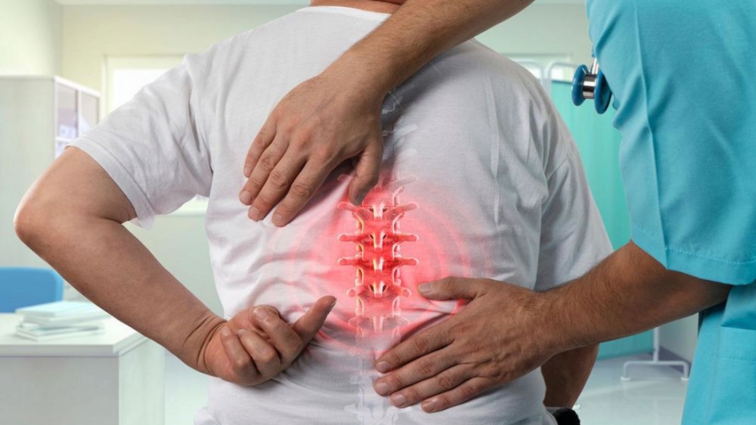
bone cyst It is usually painless, but pain may occur as the cyst grows or if an infection occurs in the cyst. There may be swelling and tenderness in the affected area. It can cause bone weakening and lead to bone fractures. If it is located near the joint, limitation and stiffness in joint movements may occur. In children, spontaneous fractures may occur as the bone with the cyst weakens.

In children, cysts located near growth plates can affect bone growth. In rare cases, the cyst may become infected. In this case, symptoms of inflammation such as fever, redness and severe pain may be observed. It is important to consult an orthopedic specialist when these symptoms are noticed. bone cyst Appropriate treatment methods are determined according to the type and location.

What Causes Bone Cyst?

It may occur as a result of abnormal cell growth and differentiation during bone development in childhood and adolescence. Especially simple cysts (unicameral bone cysts) are usually seen in children. Impacts or microtraumas to the bone tissue disrupt the blood circulation within the bone, leading to its formation. It may be caused by blockage or damage to the blood vessels in the bone. Bacterial infections that spread to bone tissue bone cyst causes its formation.

Bone infections such as osteomyelitis are examples of this condition. Metabolic diseases or genetic mutations disrupt the normal development of bone tissue. It may pave the way for cyst formation. Some cysts may form for no apparent reason. The causes of such cysts are not fully understood. Diagnosis is usually made with x-rays, MRI or CT scans. Treatment options vary depending on the type, size and location of the cyst.

What are the Bone Cyst Treatment Methods?

What is a Bone Cyst?Small ones that do not cause symptoms bone cyst It is usually monitored regularly. During this follow-up period, it is observed whether the cyst grows or causes symptoms. Painkillers and anti-inflammatory medications can be used to control pain and inflammation. In case of infection, antibiotics may be prescribed.

The contents of the cyst can be drained and sclerosing agents can be injected into the cyst cavity. This method is generally less invasive. In some cases, it may help the cyst shrink or disappear completely. Removing the cyst wall and filling the cyst cavity with synthetic materials is a common method of treatment.

In larger or aggressive cysts, complete removal (resection) of the cyst may be necessary. In this case, the missing bone tissue can be reconstructed with bone grafting. In some rare cases, especially for cysts with malignant potential, radiotherapy may be used. This treatment method uses high-energy rays to kill cyst cells or stop their growth. If the cyst is located in an intra-articular area, the cyst can be drained or removed with arthroscopic surgery. This method may be preferred because it is less invasive and offers a faster recovery process. bone cyst The most appropriate method of treatment is determined by taking into account the patient's condition.

Hammer Finger Disease

Hammer Finger Disease

It is an injury or deformation that occurs in the end joint of the finger. hammer toe disease It usually occurs as a result of excessive bending of the finger or direct blow. In this case, the tendon at the tip of the finger ruptures and the finger remains bent. If left untreated, permanent deformation may occur. Treatment may include immobilization of the finger or surgery.

What are the Symptoms of Hammer Finger Disease?

Hammer Finger Disease 
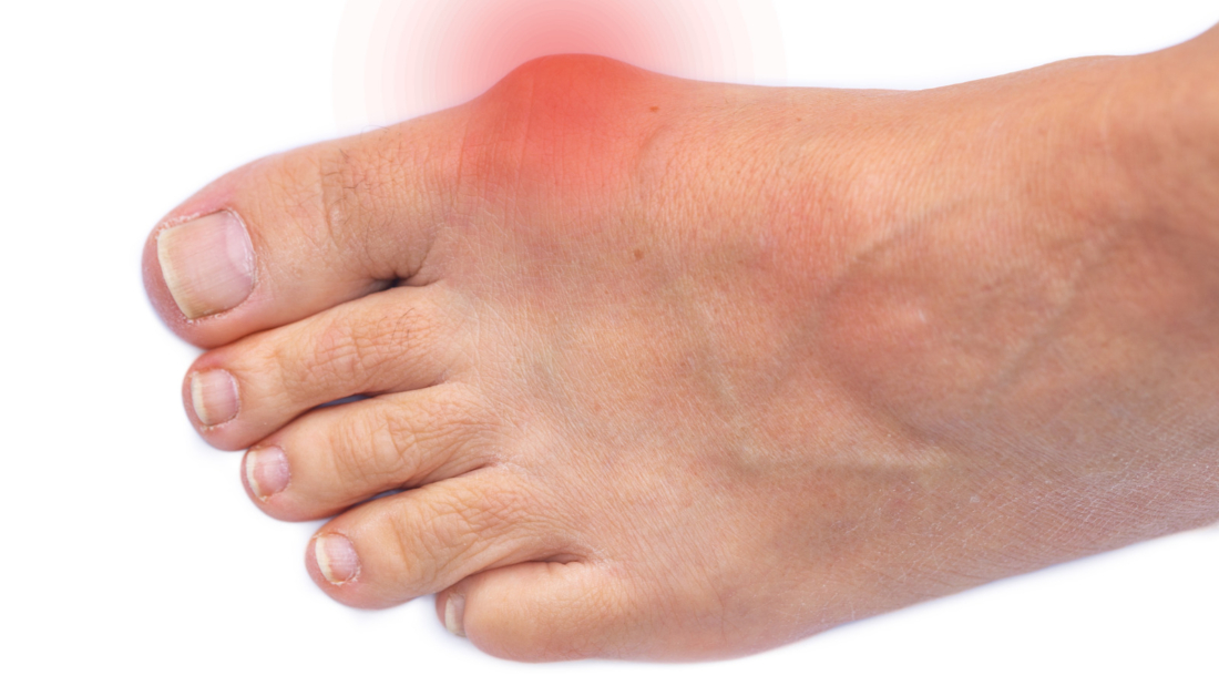
The most obvious symptom is the bending of the finger downwards from its middle joint. It causes the tip of the finger to point towards the ground. There may be pain and tenderness in the finger and joint. Pain often increases during movement or when wearing shoes. Swelling and redness may occur in the affected area. It develops due to inflammation and irritation. hammer toe disease Calluses and hardening may occur in the affected area. This occurs due to constant friction and pressure. There may be limitation and stiffness in finger movements.

This condition prevents normal movements of the finger and makes daily activities difficult. It may cause difficulties in wearing shoes. Discomfort and pain may increase due to shoes putting pressure on the toe. These symptoms hammer finger disease may indicate its existence. If any of these symptoms are present, it is important to consult an orthopedist or podiatrist. Symptoms can be alleviated with early diagnosis and appropriate treatment. Quality of life can be improved by correcting finger deformities.

What Causes Hammer Finger Disease?

In sports such as basketball and volleyball, the extensor tendon is torn when the ball hits the tip of the finger directly. Home accidents, such as a fingertip getting caught in a door or being crushed by a heavy object, cause a tear in the tendon. The tendon may be damaged or ruptured when the fingertip hits a hard surface. In some cases, tendons may weaken due to aging or joint diseases. This may cause them to get injured more easily. Treatment usually involves immobilizing the finger with a splint, sometimes surgical intervention. Full recovery can be achieved with early diagnosis and appropriate treatment.

What are the Treatment Methods for Hammer Finger Disease?

Hammer Finger DiseaseApplying a cold compress to the finger after an injury can reduce swelling and relieve pain. The finger is fixed with a splint or splint to prevent movement. The healing process is supported. This method is usually effective in mild cases. Exercises are performed to restore finger movements and strengthen muscles.

Medications such as ibuprofen naproxen are used to reduce pain and inflammation. In case of severe pain and inflammation, corticosteroid injections may be administered. If there is serious deformation in the finger joint, the bones are brought to the appropriate position with closed reduction. It is fixed with small pins. In more complex cases, joints and tendons are repaired using open surgery.

This method is generally preferred when conservative and medical treatments are not sufficient. It is important to have regular follow-up under the supervision of a doctor during the treatment process. Finger function is regained through a post-surgical rehabilitation program. hammer toe disease Early intervention and treatment methods should be applied in its treatment. It is of great importance in improving the patient's quality of life. During the treatment process, doctor's recommendations must be followed. Regular performance of the specified exercises will accelerate the healing process.

What is Kneecap Dislocation?

What is Kneecap Dislocation?

It is a condition where the patella slips from its normal place. Knee cap dislocation It occurs as a result of disruption of the path followed by the patella during movement of the knee joint. The patella may slip from its normal position, affecting the functionality of the knee. It can cause pain, a feeling of instability, and limited movement in the knee. Treatment may often include physical therapy, exercises, and rarely surgery.

What are the symptoms of kneecap dislocation?

What is Kneecap Dislocation?Severe and sudden pain may be felt. The dislocated patella usually slides out of the knee, and in this case, swelling may occur in the knee. The swelling may be tender to the touch. The normal stability of the knee may decrease. In this case, there may be a slipping or rotating feeling in the knee. The dislocated patella may slide out of the knee or rotate and change position. Knee cap dislocation When this happens, sometimes the knee can lock and become blocked when moving.

The normal strength of the knee may decrease and in this case, weakness may be felt in the knee. A crackling or popping sound may be heard in the knee. If dislocation is suspected, it is important to consult a doctor to evaluate the condition. Treatment may include repositioning or stabilizing the patella, and physical therapy may be required.

What Causes Knee Cap Dislocation?

It is possible for it to dislocate as a result of a blow to the knee area or an accident. Some people are born kneecap dislocation They may be prone to occur. This condition may result from a congenital defect in the anatomical structure of the patella. When the muscles around the knee are not strong enough and do not work evenly, patellar stability decreases. The risk may increase. Overload, sudden turns, or movements that exceed the normal range of motion of the knee may contribute.

Defects or abrasions on the cartilage surface under the kneecap can affect the stability of the patella. It may pave the way for dislocation formation. In rare cases, certain congenital conditions or abnormal anatomical structures may cause it. Knee cap dislocation People who experience knee pain often experience symptoms such as swelling and loss of stability. Treatment often varies depending on the severity of the condition and its causes. It may include conservative methods (physical therapy, exercise programs) or surgical intervention.

What are the Treatment Methods for Kneecap Dislocation?

What is Kneecap Dislocation?In mild cases, the doctor may manually push the kneecap into place. This is usually done under local anesthesia. Knee cap dislocation It can often occur due to muscle imbalance or weak muscles. Physical therapy and exercise programs are important. These programs can help prevent recurrence of dislocations by strengthening muscles and increasing flexibility. Knee bandages or special devices can be used to protect the knee during return to sports or activity. Nonsteroidal anti-inflammatory drugs are used to control pain and inflammation. Additionally, your doctor may recommend muscle relaxants or painkillers if deemed necessary.

In rare cases or when other treatment methods fail, surgical intervention is required. In this case, supportive surgical procedures may be performed to keep the patella in place. The treatment process usually ends with a plan for rehabilitation and return to activity. Physiotherapists often develop a personalized rehabilitation program. It allows the patient to return to normal activities. Knee cap dislocation The most appropriate method of treatment is determined depending on the person's condition and symptoms. Therefore, it is important to see a specialist when you have symptoms.

Tennis Elbow

What is Tennis Elbow?

It is a condition that occurs when the tendons outside the elbow are damaged as a result of overuse of the forearm muscles. tennis elbowIt is seen in people who make repetitive hand and wrist movements and takes its name from the fact that it is frequently seen in tennis players. However, it can also be common in people who do not play tennis.

What Causes Tennis Elbow?

Tennis Elbow

It occurs when the tendons outside the elbow become inflamed and damaged as a result of overuse of the forearm muscles. It develops in people who make repetitive hand and wrist movements and takes its name from the fact that it is frequently seen in tennis players.

It is usually caused by repetitive hand and wrist movements. These movements cause small tears and inflammation in the tendons. Sports such as tennis, squash and badminton can cause this condition. Especially the use of incorrect technique increases the risk of such repetitive movements.

Excessive use puts excessive stress on the tendons and causes them to wear out over time. This condition is associated with activities that require intense use of forearm muscles. It is frequently seen in tennis players. However, activities such as golf, boating, and even gardening can also cause this disorder.

Using incorrect technique during sports or repetitive tasks increases the stress on the tendons. Additionally, the use of improper equipment can also cause tendons to become overloaded.

With the aging process, the flexibility and durability of tendons decrease. Therefore, the risk is higher in individuals between the ages of 30-50. Additionally, genetic predisposition may also play a role in the development of this disorder.

Weak forearm muscles and inadequate conditioning cause the tendons to be more sensitive to loading. Lack of exercises that increase muscle strength and endurance may increase your risk.

As a result, it is caused by repetitive motion, overuse, etc. Being aware of these risk factors and taking precautions plays an important role in preventing them.

What are the symptoms of tennis elbow?

tennis elbowIt is a condition that occurs when the tendons outside the elbow become inflamed and damaged. It usually develops as a result of repetitive hand and wrist movements.

  • The most common symptom is pain and tenderness felt on the outside of the elbow. This pain can radiate from the elbow to the forearm and usually starts mild and becomes severe over time. Sensitivity increases when the elbow area is touched or pressure is applied.
  • People may experience weakness and loss of strength, especially in activities that require the use of hands and wrists. This condition manifests itself as difficulty grasping objects, not being able to hold them tightly, or difficulty doing simple tasks.
  • Pain may increase during hand and wrist movements such as shaking hands, lifting something, bending or rotating the wrist. Pain becomes more pronounced with repetitive movements or certain positions.
  • It can make daily activities difficult. For example, even simple tasks such as lifting a cup, writing, brushing your teeth, or turning a doorknob can become painful. This condition can significantly reduce quality of life.
  • Pain usually subsides after rest but reoccurs when activities resume. In advanced cases, pain may continue at rest.

Symptoms appear in various ways. If you are experiencing these symptoms, you should consult a healthcare professional for proper diagnosis and treatment.

Tennis Elbow Treatment

Tennis Elbow

tennis elbowIt is a condition that causes pain and tenderness on the outer part of the elbow. Treatment of this condition is done using various methods to relieve pain and promote healing of the elbow.

The first step of treatment is to rest the elbow and avoid activities that cause pain. It is important to protect the tendon by avoiding repetitive movements and heavy lifting. Choosing alternative activities can speed up the recovery process.

Ice can be applied to the elbow to reduce pain and swelling. Wrapping ice in a thin towel and applying it for 15-20 minutes several times a day can be effective. NSAIDs can be used to relieve pain and inflammation. These drugs should be taken with the recommendation of a doctor.

Physical therapy offers specific exercise programs to strengthen elbow muscles and increase flexibility. Exercises determined by a physical therapist can help heal the tendon.

Special elbow bands or wristbands protect the tendon by reducing stress on the elbow. These supports can provide relief to the elbow during daily activities. In cases of severe pain, corticosteroid injections relieve inflammation and pain. However, this treatment method is not recommended as a long-term solution and should be applied in limited numbers.-

When conservative treatment methods fail, surgical intervention may be considered. Surgery is performed to repair or remove damaged tendon tissue. This method is used as a last resort and the recovery time is longer.

tennis elbow Treatment is determined according to the patient's condition and the severity of the symptoms. With early diagnosis and appropriate treatment, most patients can recover without the need for surgical intervention. It is important to follow your doctor's recommendations during the treatment process.

Torus Fracture

What is a Torus Fracture?

It is a type of bone damage seen in children in which the bones are not completely broken, only one side is bent inward. It occurs at the ends of long bones, radius and ulna bones. torus fractureIt is more common in children than in adults because their bones are not yet fully hardened and flexible.

What Causes Torus Fracture?

Torus Fracture

It is a type of bone damage that is more common in children. These fractures usually occur in younger patients, where the bones are flexible and can bend rather than break completely. Torus fractures occur because bones become softer and more flexible during the growth and development process.

Falls experienced by children while playing or doing sports are among the most common causes. During a fall, the sudden force applied to the bone causes one side of the bone to collapse inward. Thus, a fracture occurs in the torus.

Because children are active, they can be exposed to direct impacts. For example, events such as being hit by a ball or colliding with another child can lead to torus fractures, especially in the arms and legs.

Sudden and violent bending can also cause torus fractures. This type of strain occurs especially in long bones (for example, in the wrist or arm). Constant and repeated stress can slowly build up on bones, causing torus fractures. These types of fractures are generally not sudden, but damage that develops over time.

Children's bones are more flexible and less mineralized than those of adults. Therefore, it can occur even with little strain. Children's active lifestyle and constant change of bone structure increase the risk factors for such fractures.

When a torus fracture is diagnosed, treatment usually includes immobilization with a cast or splint. Because children's bones heal quickly, torus fractures heal without complications or permanent damage.

What are the Symptoms of Torus Fracture?

Torus fracture is a type of bone damage seen especially in children and is characterized by a part of the bone collapsing inward. Because children's bones are more flexible than those of adults, these types of fractures often occur during childhood.

  • Significant pain is felt, especially when the fracture area is touched or moved. Pain may prevent the child from using or pressing on that area.
  • Swelling occurs in the fractured area. This swelling occurs when fluid and inflammatory cells accumulate around the fracture and usually occurs shortly after the fracture.
  • The fracture area may be extremely tender to the touch. This sensitivity is caused by irritation of the nerve endings in the area of the fracture.
  • A typical symptom is the inability to move or use the fractured area normally.
  • Sometimes, bruising or bleeding under the skin may occur in the fracture area. This may vary depending on the severity of the impact and the location of the fracture.
  • There is usually no obvious deformity in torus fractures. However, in some cases, a slight deformity may be noticed in the fracture area.

If the child has any of the symptoms, it is important to seek medical help immediately. Early diagnosis and treatment can help the child recover faster and prevent possible long-term damage. In cases of suspected fracture, the doctor makes a definitive diagnosis by taking an x-ray and determines the appropriate treatment method.

Torus Fracture Treatment Methods

Torus Fracture

Torus fracture is a type of fracture, especially seen in children, in which one side of the bone collapses inward and the other side remains intact. These types of fractures are generally considered less serious than complete fractures because of children's flexible bone structure. Treatment methods focus on the child's capacity for rapid recovery.

The broken arm or leg can be put in a cast or, as a more flexible option, a splint can be used. This method is necessary to ensure that the bone heals in the correct position and reduces pain.

The pain and discomfort caused by the fracture can be controlled with painkillers recommended by the doctor. It is important not to put any weight on the fractured area during healing. The child should not engage in physical activities.

Adequate rest is critical for rapid recovery. During the first few days, it is recommended that the child get as much rest as possible. The healing process of the fracture should be monitored with regular doctor checks. During these checks, the condition of the cast or splint is checked. If necessary, it is replaced with a new one. Additionally, the healing status of the fracture is evaluated.

At the end of the healing process, an x-ray may be taken to confirm whether the fracture has healed completely. After immobilization, physical therapy is required to regain mobility and strength in the fracture area. This is especially important after long-term immobilization.

Torus fractures are conditions that do not cause serious complications and heal completely with good treatment. Compliance with the doctor's recommendations and regular check-ups ensure that the child has a rapid recovery process.